Few patients enjoy having their blood drawn or getting an IV insertion, with some even dreading these common procedures. Venipuncture can be especially unpleasant when a suitable vein is difficult to find, and the patient is pricked and poked many times before success.
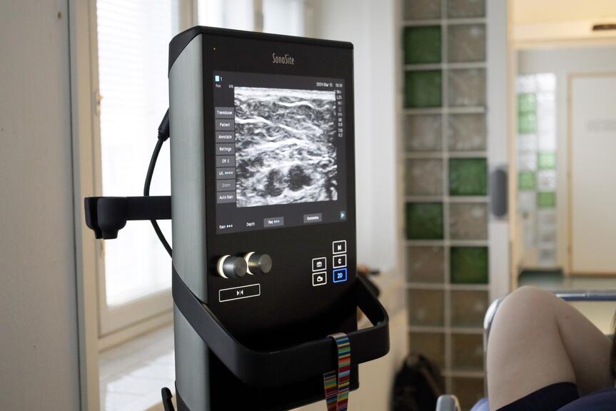
To help with these procedures, Laakso Hospital is using an ultrasound device that makes it so easy to guide a needle into a suitable vein that the first-attempt success rate is as high as 98% – making the process almost painless for the patient.
“Without ultrasound, needle insertion is practically ‘blind’ – deep veins are impossible to see, and even superficial veins can be difficult to detect if the patient has a lot of tattoos, for example,” says Sari Roos, Nursing Instructor at Laakso Hospital.
Roos has been working as a nursing instructor at Laakso Hospital for 16 years and has seen the benefits of the ultrasound device first-hand. The device has been in use for four years now, with Roos teaching nurses how to operate it.
When you can see a patient’s vein on a monitor, choosing the right size of needle becomes easy. For example, nurses can use a needle that is thinner and longer than normal to access particularly deep veins, with the patient feeling little to nothing at all.
“The device also improves patient safety by showing us the locations of nerves, tendons and different types of blood vessels. For example, arteries should never be punctured; the device helps us to easily differentiate between veins and arteries,” Roos clarifies.
An added benefit of using ultrasound imaging is that sometimes blood clots can be detected in the patient’s arm while performing a procedure. Such findings are always reported to the attending physician, who can then assess follow-up procedures.
From enhanced patient outcomes to clear cost savings
The ultrasound device used at Laakso Hospital also saves hundreds of thousands of euros per year due to patients not having to be sent to specialised health care, which is always subject to a procedure fee and an ambulance fee.
“In 2023, the team performed a total of 598 venipunctures on the patients of Laakso Hospital alone. These were all cases where the procedure could not have been performed without the ultrasound device. Without it, patients would have had to be referred to specialised health care, or their treatment plans would have had to be changed,” Roos says.
In addition to saving money, the device also saves time. For example, nursing staff can start administering intravenous medication more quickly when the patient does not need to be transferred to specialised health care to get access to a vein. Transfers are also always taxing for the patient, especially if they are in poor condition.
“At Laakso Hospital, we have trained a team of nurses to use the ultrasound device so that they can rapidly respond to the needs of our different wards. Learning to use the ultrasound device is easy and has provided us with tremendous benefits,” Roos sums up.


